

The device is intended for imaging and diagnostics of pathological changes of the breast tissues.
The electrical impedance method of diagnostics is based on the fact that electrical conductivity (ability to conduct electrical current) of biological tissues correlates considerably to their physiological status. It is well-known that many tumours, in particular malignant breast tumours, feature electrical conductivity, which differs significantly from the electrical conductivity of surrounding healthy tissues.
This phenomenon explains high sensitivity of the mammograph. The device enables, using the Electrical Impedance Tomography (EIT) method, to obtain the picture of biological tissues electrical conductivity distribution in the cros-sections of the breast at different frequencies. Distribution of electrical conductivity in every cross-section is visualized on a PC screen. The key advantages of electrical impedance method of diagnostic are as follows: absolute safety of examination, high level of information content, compactness, affordability and simplicity of the examination procedure.
The mammograph is intended for usage in conditions of specialized branches of hospitals and clinics. The device software supports exchange of data and images in DICOM format.
PRINCIPLE OF THE DEVICE OPERATION
Various organs and tissues in a human body possess different electrical properties. For instance, it is a well-known fact that many tumours, in particular malignant breast tumours, feature electrical conductivity (ability to conduct electrical current), which differs considerably from the electrical conductivity of surrounding healthy tissues. The MEM makes it possible to obtain the picture of biological tissues electrical conductivity distribution in the breast in its transverse sections and locate such tumours in the obtained images.
The multifrequency electrical impedance mammograph (MEM) principle of operation is based on the method of electrical impedance tomography (EIT). The present device has come to replace the previously developed single-frequency device MEIK. Scientific research in the field of electrical impedance tomography has been developing since the middle of 1980s. The method enables the examiner, utilizing a full (in mathematical sense) set of electrical measurements, performed with the help of a multielectrode system, to reconstruct spatial distribution of electrical properties inside an object. The process of reconstruction is carried out through solving of the so-called inverse problem for the equation of electric field in inhomogeneous medium. A group of Russian scientists, comprising researchers from the Institute of Radio-engineering and Electronics of the Russian Academy of Sciences, is one of the world leader in the field of the EIT.
Below comes a list of some publications of the abovementioned researchers in scientific journals:
- Korjenevsky, et al “Electrical impedance computerized tomograph for medical applications”, Instruments and Experimental Techniques, v. 40, No 3, p. 415-421 , 1997;
- V. Cherepenin, et al “A 3D electrical impedance tomography (EIT) system for breast cancer detection”, Physiol. Meas., v. 22(1), p. 9-18, 2001;
- V. Cherepenin, et al, “Three-dimensional EIT imaging of breast tissues: system design and clinical testing”, IEEE Trans. Medical Imaging, v. 21(6), p. 662-667, 2002;
- Korjenevsky A.V. et al, “Electrical impedance tomography system for 3D imaging of the breast tissues”, Biomeditsinskie tekhnologii i radioelektronika, N 8, p. 5-10, 2003;
- Trokhanova O.V., Okhapkin M.B. and Korjenevsky A.V. “Dual-frequency electrical impedance mammography for the diagnosis of non-malignant breast disease”, Physiol. Meas., v. 29, p. S331-S344, 2008.
TECHNICAL FEATURES:
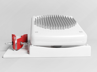
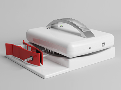
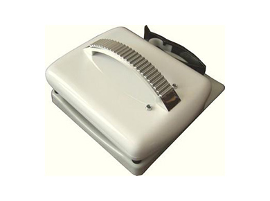
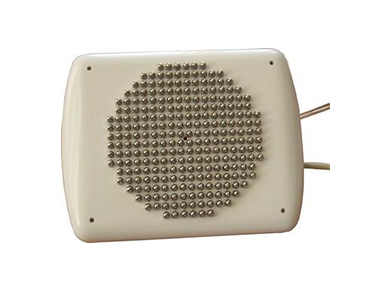
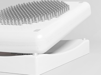
- The device comprises a measuring block with a built-in microprocessor control system, which houses a matrix with 256 electrodes.
- Two remote electrodes, which are positioned on a patients arm, are connected via a cable to the measuring block.
- During the scanning process the device, utilizing one out of 256 electrodes of the matrix sequentially injects into the patient’s body weak alternative electric current and registers corresponding distributions of potentials on its surfaces using the rest of the electrodes.
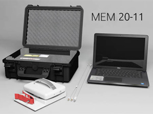
- The obtained data are used then for reconstruction of the electrical impedance images with the help of the mathematical algorithms utilizing a personal computer to which the device is connected vie the USB port.
- The utilized algorithm allows to reconstruct three-dimensional distribution of electrical conductivity (in the form of tomographic cross-sections of various depths) and to obtain highly detailed images.
- The utilized measuring scheme and the algorithm of image reconstruction make imaging results practically insusceptible to the skin surface condition.
- The MEM makes it possible to carry out examination with various frequencies, which in some cases enhances precision of diagnostics since depending on the tissues status, their electrical properties correlate differently with frequencies.
ADVANTAGES OF THE METHOD
1. Electrical impedance mammography is a simple, affordable and inexpensive method; it is radiation-free and non-invasive. In addition to visual assessment the method enables the examiner to carry out a quantitative evaluation of the mammograms, which is extremely important in differential diagnostics of various breasts states and their pathologies.
2. The method has no contraindications and can be used as often as requited which is very important for case monitoring of women with breast pathologies as well as for case management, assigning of combined oral contraceptives or hormone replacements therapy.
3. It allows to examine pregnant women and puerpera.
4. The electrical impedance mammograph can be used in inpatient and outpatient departments of hospitals, maternity welfare centres, doctors’ offices and other institutions of public health. Thanks to its portability and compactness the device can be used for breast examination in remote areas, which lack stationary equipment.
5. The method of multifrequency electrical impedance mammography significantly improves diagnostic results of dyshormonal diseases. Compared with the single-frequency electrical impedance mammography, multi-frequency mammography allows not only to diagnose mastopathy, but to differentiate a cystic mastopathy from an acystic one, thus, defining a risk group of patients with possible development of breast cancer for a closer examination and supervision.
6. The multi-frequency method of electrical impedance mammography makes it possible to diagnose changes in tissue status at mastalgia, which other methods of breast examination can not provide.
7. The multi-frequency method of electrical impedance is very effective in diagnostics of benign and malignant tumours of breast. Its sensitivity (the number of patients in whom the disease symptom is detected or symptom rate) is not below 76% and specificity (absence of symptom rate in case of healthy people) is 75%. Compared with the method of X-ray mammography, the latter is characterized by the sensitivity 71% – 87% and specificity – 38%.